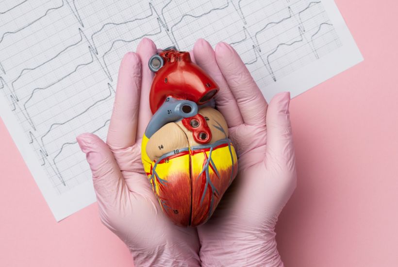
The heart has two upper receiving chambers, called atria, and two lower pumping
chambers, called ventricles. The heart also has a built-in electrical system that controls and coordinates its pumping function.
A heartbeat is caused by an electrical impulse traveling through an intricate system of electrical pathways in the cells of the heart tissue. The impulse travels through the upper and then lower chambers, causing the alternate contraction and relaxation that results in pumping of blood.
The impulse originates in the sinus (sinoatrial, or S-A) node, the heart’s natural pacemaker located within muscle at the top of the right upper atrium, and travels across both atria. Then the impulse collects, or pauses, in the atrioventricular (A-V) node, located within muscle near the Centre of the heart, and travels down the ventricles. The A-V node is the only normal electrical connection between the atria and the ventricles. At 3 to 4 millimeters, the sinus and A-V nodes are tiny, the size of a few grains of sugar.
Heart block occurs when there is a delay in the conduction of the electrical impulse through the heart. In most cases, heart block is caused by a problem with the A-V node.
There are several types of heart block, including:
Complete heart block: The most common type of heart block in children is complete heart block, also called third-degree heart block. In complete heart block, the electrical impulse never gets past the A-V node. The only reason a person can survive is that another, weaker natural pacemaker takes over in the ventricles. The ventricles are able to pump blood out to the body, but more slowly than normal.
First-degree heart block: In first-degree heart block, the electrical impulses move through the A-V node more slowly than usual.
Second-degree heart block: In second-degree heart block, there is a delay in the electrical impulse reaching the ventricles. Skipped heartbeats and a slower-than-normal heart rate may result. There are two types of second-degree heart block. In some cases, second-degree heart block will eventually progress to complete heart block.
Bundle branch block: There is a flaw in the bundle branches, the pathways for the electrical impulse running along the right and left ventricles. This can be congenital or acquired, and is often seen after surgery to close a defect in the septum (the wall of tissue) between the ventricles.
Symptoms of complete heart block can include:
The other types of heart block in children can also cause these symptoms, though they are usually less severe.
In infants born with complete heart block, symptoms can also include:
Sometimes heart block may not cause any symptoms. In rare cases, complete heart block or second-degree heart block can cause sudden death, even in a person with few or no symptoms.
Sometimes complete heart block is diagnosed prenatally. With fetal echocardiography, doctors may notice a difference between the rate at which the upper and the lower chambers are beating. Babies born with complete heart block can have wires for a temporary pacemaker placed within minutes.
In other cases, heart block in children is not diagnosed until a later age, or even adulthood.
Diagnosis of heart block may require some or all of these tests:
In many cases, complete heart block will eventually require a pacemaker. This is a battery-operated device that doctors implant under the skin. Leads (wires) attached to the device are placed on the surface of the heart (in infants), or are threaded through veins directly into the heart (in older children and teenagers). The pacemaker wires serve as replacement sinus and A-V nodes, and the electrical signals they send reach both the atria and the ventricles, correcting the heart block.
Implanting and maintaining a pacemaker comes with some risk; doctors factor this into their decision about what treatment to pursue. Doctors will carefully monitor these patients so they know if there is a change in the patient’s condition and a pacemaker should be placed.
In some cases of complete heart block, the heart rate is sufficient and a pacemaker won’t be necessary. Rarely, second-degree heart block will require a pacemaker. First-degree heart block and bundle branch block usually don’t require any treatment.
Children with complete heart block will require lifelong care by a cardiologist. Those with the less severe forms of heart block should also continue to see a cardiologist regularly.
Children with pacemakers can lead physically active and healthy lives. However, some sports and activities may not be allowed.
